
Advances in bronchoscopy over the past several years have resulted in an array of advanced diagnostic and treatment modalities now available to pulmonologists; however, the roles of each modality and the consensus on when and how to use them are still evolving.
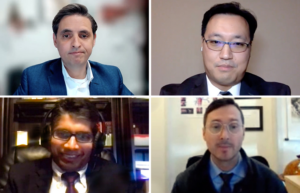
During a session on Tuesday, Diagnostic and Therapeutic Bronchoscopy: Year in Review, a panel of expert pulmonologists reviewed recent research findings in hot topics such as bronchoscopy for lung nodule diagnosis, endobronchial ultrasound (EBUS), lung cryobiopsy, and bronchoscopic lung volume reduction (BLVR).
Francisco Almeida, MD, MS, FCCP, Professor of Medicine in the Department of Pulmonary, Allergy, and Critical Care at the Cleveland Clinic, began the session with a review of recent literature on EBUS, including the use of EBUS-guided transbronchial fine needle aspiration (EBUS-TBNA) for non-small cell lung cancer (NSCLC) staging.
“This is probably the area where we see the most data coming out today,” Dr. Almeida said.
He cited findings from a recent study conducted across seven centers in the United Kingdom and the United States that evaluated EBUS-TBNA performance for PD-L1 assessment and reported an overall adequacy rate of approximately 95%. However, he noted, the findings from a study conducted by a group in the Netherlands highlighted important false-negative concerns.
“They reported quite a bit of false-negative PD-L1 staining, up to 36%, when the sample is immersed directly in ethanol fixative instead of directly into formalin,” Dr. Almeida said. “Our practice at Cleveland Clinic is to collect a few passes and place these directly into formalin for PD-L1, and our adequacy numbers are pretty consistently in the mid- to high-90 percentile.”
He referred to an ongoing registry in the UK that collects EBUS performance data and, as research in this area continues to evolve, he believes a similar initiative would be of great value in the US.
“The quality numbers that they demonstrated are pretty impressive. They can look at their biomarker data to evaluate where the problem is and try to work on getting it better,” Dr. Almeida said. “I believe it is time that we in the US and other countries copy our British colleagues by doing the same and try to create more uniform data assessment for the procedures we do.”
In the next presentation, Satish Kalanjeri, MBBS, FCCP, Associate Professor of Medicine at the University of Missouri-Columbia and Chief of Interventional Pulmonology at Harry S. Truman VA Hospital, discussed recent evidence for guided bronchoscopy for peripheral lung nodules, focusing on diagnostic yield as the key metric.
He reviewed recent data on the use of Lung Vision, an AI-based platform that uses augmented fluoroscopy to guide navigation to the lesion, as both a standalone modality and paired with adjunct cone-beam computed tomography (CBCT).
A multicenter prospective study led by investigators at the Cleveland Clinic examined navigational success as defined by radial EBUS showing a signal for the target lesion with diagnostic yield as the other primary endpoint.
“Although the mean lesion size was just under 3 cm for each lesion, there was a pretty significant range—up to 80 cm,” Dr. Kalanjeri said. “Navigational success was quite high, almost 93%, the overall diagnostic yield was about 75%, and if you take out the larger lesions and only analyzed the lesions under 3 cm, the diagnostic yield fell to about 69%, which is still pretty good.”
A separate single-center observational study published earlier this year using the same endpoints looked at the use of CBCT as an adjunct to the Lung Vision platform.
“This study reported a diagnostic yield of 71% after the procedure, which increased to 88% at the end of 1 year,” Dr. Kalanjeri said. “Looking at this and data from other studies demonstrates that these newer technologies, when they have cone-beam CT scan or augmented fluoroscopy as adjuncts, do offer some diagnostic yield advantage.”
Jeffrey Thiboutot, MD, Assistant Professor of Medicine, Division of Interventional Pulmonology, Pulmonary and Critical Care, at Johns Hopkins University, followed with a review of recent findings in cryobiopsy and therapeutic bronchoscopy.
Among the studies he reviewed was the COLDICE trial, which evaluated the agreement between concurrent cryobiopsy and surgical lung biopsy from the same patients for both histopathological and multidisciplinary discussion diagnoses.
“This is one of the biggest studies to come out in the cryobiopsy realm in the past 10 years or so,” Dr. Thiboutot said. “It was a really well-done trial performed at nine centers in Australia where they enrolled 65 patients who needed a histopathologic diagnosis to aid in securing a diagnosis of interstitial lung disease.”
The results, he said, showed high concordance in both the histopathologic interpretation and the multidisciplinary diagnosis between the cryobiopsies and the surgical biopsies.
“The backside of this, as with all cryobiopsy studies, is the safety profile, which is still an important question,” Dr. Thiboutot said. “However, I think current literature does support the integration of cryobiopsy into disease diagnostic algorithms for the workup of ILD.”
Concluding the session, George Cheng, MD, PhD, FCCP, Associate Professor of Medicine and Director of Interventional Pulmonology, Bronchoscopy, and Pleural Disease at the University of California-San Diego, discussed the outlook for bronchoscopic therapies in peripheral lung cancer.
“Peripheral bronchoscopy-guided therapeutic options are emerging, but there are major clinical questions that need to be addressed—how effective it is, how it compares to standard care, and how it fits within the therapeutic algorithm,” Dr. Cheng said. “But suffice it to say that this is a very exciting time for the field of bronchoscopy, where we are on the doorstep to deliver diagnosis, staging, and treatment in one event.”
Missed this session? Watch the recording in the “Live Session Recordings” tab of the On Demand section in the meeting platform. Recordings will be available approximately 48 hours after the original session time.
NOW IN SESSION
CHEST 2021 • OCTOBER 17-20 • INFORM. INSPIRE. INNOVATE.
Immerse yourself in the experience of CHEST 2021! Your registration includes hundreds of sessions presented by our top faculty and featured speakers, live discussions with the experts, interactive gaming, and much more. Even better, you’ll have access to everything (including post-meeting bonus content) until October 2022!
Haven’t registered yet? It’s not too late. Register now for full access.